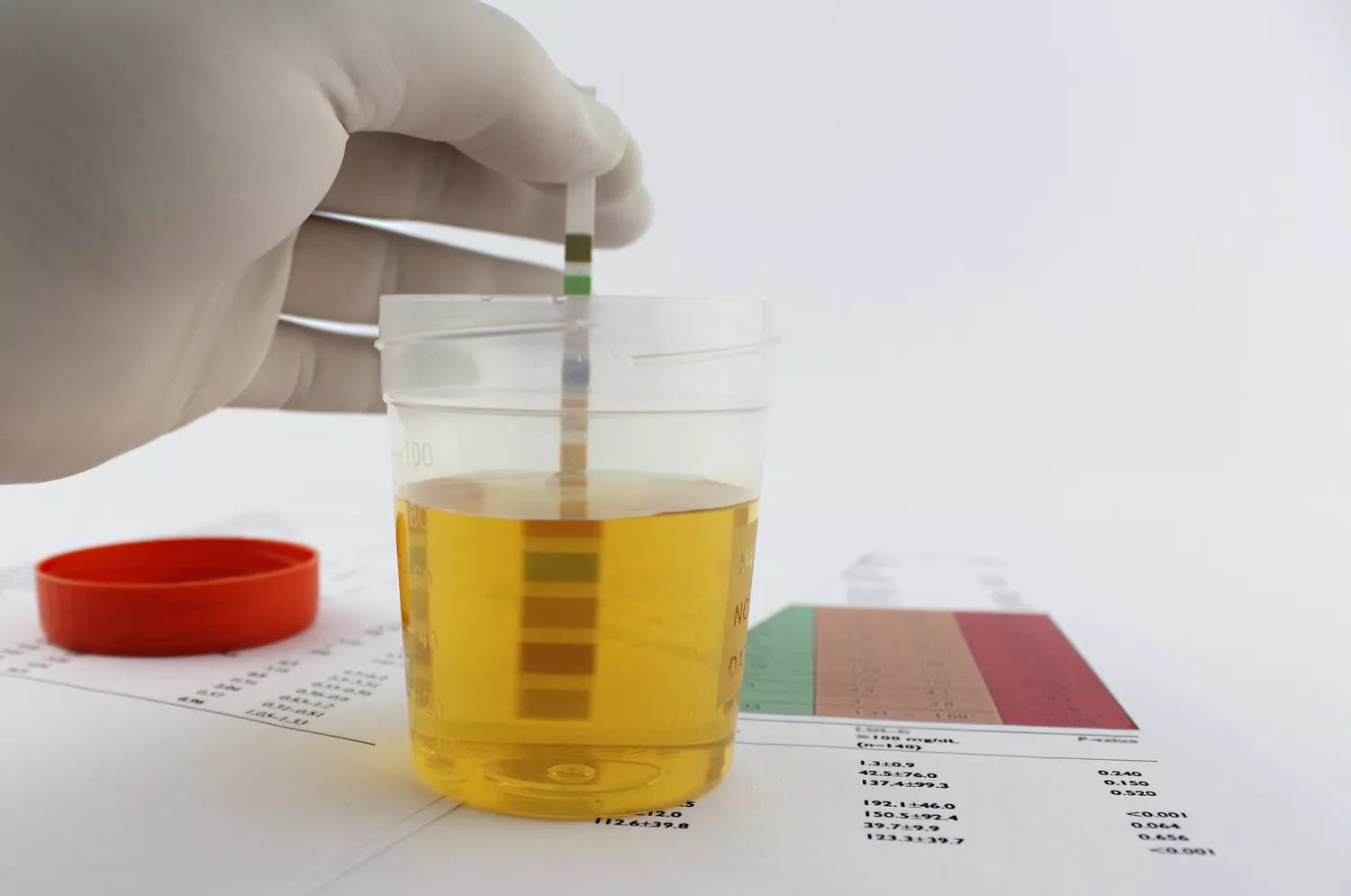
The presence of protein in urine, a condition medically known as proteinuria or sometimes albuminuria, is one of those clinical findings that often causes immediate concern, yet it is simultaneously one of the most frequently misunderstood diagnostic results. While trace amounts of protein can sometimes be a transient, benign finding related to exercise, fever, or even stress, its persistent or significant presence is a critical alarm bell. The kidneys, acting as the body’s sophisticated filtration system, are designed to retain larger molecules like proteins in the bloodstream, allowing only waste products to pass into the urine. Therefore, detecting protein in the final filtrate suggests a fundamental issue with this delicate filtering process. This issue can range from temporary physiological stress to chronic, silent damage within the renal structures. Understanding what these results truly signify requires moving beyond simple definitions and appreciating the intricate anatomy and underlying conditions involved. The following examination delves into the various reasons why this essential nutrient might be escaping the circulation, focusing on the nuanced distinctions between temporary overflow, functional changes, and genuine pathology.
The Kidney’s Filtration Process is Not Flawless Under Stress
To appreciate why protein leaks into the urine, one must first grasp the highly specialized filtration apparatus within the kidney: the glomerulus. This intricate network of capillaries functions as a selective barrier, allowing water and small solutes to pass through, while larger plasma proteins, particularly albumin, are intended to be kept within the bloodstream. However, this kidney’s filtration process is not flawless under stress. Certain conditions, even seemingly innocuous ones, can temporarily compromise this barrier’s integrity. For instance, a phenomenon known as orthostatic proteinuria occurs when protein is present in the urine only when the person is upright and active, disappearing when they lie down. Similarly, an acute, significant fever or an episode of intense, prolonged physical exertion can cause a transient increase in urine protein. These are often categorized as functional or transient proteinuria, where the underlying kidney structure is sound, but the high-demand state momentarily alters the pressure dynamics or permeability. These temporary leaks are typically not indicative of chronic disease and usually resolve spontaneously, yet they underscore the fragility of the glomerular filter when subjected to systemic perturbation. The challenge lies in distinguishing these fleeting anomalies from persistent, disease-driven leakage.
Persistent Leakage is a Red Flag for Glomerular Barrier Integrity
When protein is detected consistently across multiple urine samples, the possibility of a systemic or localized pathological process becomes much more substantial. Persistent leakage is a red flag for glomerular barrier integrity, signaling damage to the structures responsible for keeping proteins in the blood. The vast majority of clinically significant proteinuria originates from this damage. Conditions that cause this sustained breakdown include diabetic nephropathy, the most common cause in the developed world, where chronically elevated blood glucose damages the tiny blood vessels over time. Another major culprit is hypertension, or high blood pressure, which physically stresses the glomerular capillaries, leading to thickening and scarring. Furthermore, primary kidney diseases, collectively termed glomerulonephritis, involve various inflammatory and non-inflammatory processes that directly assault the filtering units. This persistent presence of protein, especially albumin, is not just a symptom; it’s also believed to contribute to the progression of kidney injury by overloading the tubular system, creating a vicious cycle of destruction and leakage.
Diabetic Nephropathy is the Most Common Etiology in Many Regions
The insidious progression of diabetic nephropathy is the most common etiology in many regions for chronic kidney disease, and its early sign is microalbuminuria, a small but persistent leak of albumin. Uncontrolled or poorly managed blood sugar levels lead to the accumulation of advanced glycation end-products (AGEs) and structural changes within the glomerulus. Specifically, the podocytes, which are highly specialized cells that act as the final, fine-tuned filtration sieve, are damaged. The thickening of the glomerular basement membrane and loss of these podocytes progressively widen the pores of the filter, allowing more and more albumin to escape. This leakage often begins years before a noticeable decline in the kidney’s overall filtration rate, making it a crucial marker for early intervention. The shift from microalbuminuria (small amounts of albumin) to macroalbuminuria (larger amounts) signals a more advanced stage of disease where kidney function decline is generally inevitable without aggressive treatment aimed at stringent blood sugar and blood pressure control.
Certain Systemic Conditions Can Lead to Overflow Proteinuria
Beyond issues with the filter itself, certain systemic conditions can lead to overflow proteinuria due to an overproduction of certain proteins that saturate the kidney’s reabsorption capacity. This is distinctly different from the filtration barrier damage previously discussed. For example, conditions like multiple myeloma, a cancer of plasma cells, produce excessive amounts of light chains, which are small antibody fragments. While the glomerulus might filter these effectively, the renal tubules—the structures responsible for reabsorbing nearly all filtered protein—become overwhelmed. The tubules cannot keep pace with the massive protein load, and the excess spills into the urine. This type of proteinuria is often a critical diagnostic clue for underlying hematologic malignancies. The protein detected is typically not albumin, but rather these lighter, smaller globulins, thus requiring specific testing, often electrophoresis, to identify the precise type of protein involved and pinpoint the systemic disease responsible for its overabundance in the circulation.
The Ratio of Albumin to Creatinine is Often Utilized
In modern clinical practice, a single measurement of protein concentration in a random urine sample is considered less reliable due to variations in fluid intake and urine concentration. For this reason, the ratio of albumin to creatinine is often utilized as a standardized and more accurate way to quantify proteinuria. Creatinine, a waste product of muscle metabolism, is produced at a relatively constant rate and is excreted by the kidneys. By comparing the amount of albumin in the sample to the creatinine concentration (the urine albumin-to-creatinine ratio, or UACR), clinicians can normalize the result to the patient’s muscle mass and hydration status, providing a much clearer picture of the protein leak over a 24-hour period. A persistently elevated UACR is the gold standard for monitoring the progression of chronic kidney disease and assessing the efficacy of treatments, such as angiotensin-converting enzyme (ACE) inhibitors or angiotensin receptor blockers (ARBs), which are specifically prescribed to reduce the pressure within the glomerulus and decrease protein leakage.
Tubulointerstitial Diseases Affecting Reabsorption Also Play a Role
While glomerular damage accounts for the majority of severe proteinuria, tubulointerstitial diseases affecting reabsorption also play a role, contributing to what is known as tubular proteinuria. The renal tubules, which extend from the glomerulus, are critical for reabsorbing small proteins and essential substances back into the blood. Damage to the cells lining these tubules, which can occur from toxins, certain medications (like some non-steroidal anti-inflammatory drugs or certain antibiotics), or chronic infections, impairs this reabsorption function. The result is the appearance of small-molecular-weight proteins in the urine that would normally be reabsorbed. Although tubular proteinuria is typically less massive in quantity than severe glomerular proteinuria, it is equally important as a diagnostic indicator. It points towards disease processes localized primarily in the kidney’s plumbing and interstitial tissue, prompting a focused investigation into drug histories, environmental exposures, or specific inflammatory syndromes.
Proteinuria Can Occur Without Any Palpable Symptoms in the Patient
One of the most concerning aspects of this condition is that proteinuria can occur without any palpable symptoms in the patient for years, even decades. This characteristic of silent progression is precisely what makes screening for it so vital, particularly in high-risk populations like individuals with diabetes or hypertension. In the early stages of kidney damage, the body’s compensatory mechanisms, including the remaining healthy nephrons, mask the functional decline. The leakage is detected only via laboratory testing. However, once the amount of protein leakage becomes very high—a condition often termed nephrotic range proteinuria—the patient may begin to experience generalized swelling, or edema. This swelling occurs because the significant loss of albumin (the primary protein responsible for maintaining osmotic pressure) from the blood reduces the circulating fluid’s ability to draw water back from the tissues, leading to fluid accumulation, often noticeable around the eyes, ankles, and feet.
Autoimmune Disorders Can Instigate a Complex Cascade of Inflammation
Beyond diabetes and hypertension, various autoimmune disorders can instigate a complex cascade of inflammation that directly attacks the kidney’s filtering units, leading to significant proteinuria. Systemic lupus erythematosus (SLE) is a prime example, where the body’s own immune system produces antibodies that deposit within the glomerular structures, causing severe inflammation—a condition known as lupus nephritis. Similarly, conditions like vasculitis, which cause inflammation of blood vessels throughout the body, can target the renal capillaries. The resultant injury to the filtration barrier is often severe and rapid, frequently leading to the swift development of nephrotic syndrome and requiring aggressive immunosuppressive therapy to halt the destructive process. The type of protein found and the accompanying clinical features are essential clues for the nephrologist to differentiate these immune-mediated renal injuries from other, less inflammatory causes.
Genetic Predispositions That Affect the Podocyte Structure
A less common but equally important cause involves genetic predispositions that affect the podocyte structure. These are congenital conditions where the kidney’s filtering cells are structurally flawed from birth. Conditions like Focal Segmental Glomerulosclerosis (FSGS), in some of its forms, and Alport syndrome are examples where mutations in genes coding for structural proteins lead to defects in the podocytes or the glomerular basement membrane. These defects weaken the filter’s integrity, causing protein leakage that often begins in childhood or adolescence. The fact that the underlying defect is genetic means the damage is intrinsic to the kidney’s development, presenting a distinct set of challenges for long-term management. Identification of these genetic markers is increasingly important for diagnosis and prognosis, moving the field towards highly personalized treatment strategies that differ substantially from those used for acquired diseases like diabetic nephropathy.
The Therapeutic Strategy Must Be Tailored to the Specific Etiology
Finally, the treatment of proteinuria is never a one-size-fits-all approach. The therapeutic strategy must be tailored to the specific etiology and the quantity of protein leakage. If the cause is a simple overflow from multiple myeloma, the primary treatment targets the cancer, not the kidney directly. If it is due to diabetes, the focus is intense glycemic and blood pressure control. For inflammatory autoimmune conditions like lupus nephritis, the mainstay of therapy involves immunosuppressive drugs. Crucially, regardless of the underlying cause, medications that block the Renin-Angiotensin-Aldosterone System (RAAS inhibitors), such as ACE inhibitors and ARBs, are almost universally employed when persistent proteinuria is present. These drugs have the unique ability to dilate the efferent arteriole in the kidney, reducing the pressure inside the glomerulus, thereby directly lessening the mechanical force that drives protein across the damaged filter. This reduction in proteinuria is one of the most reliable predictors of long-term kidney survival.